系統名: NOD/ShiLtJGpt-Prkdcem26Cd52Il2rgem26Cd22/Gpt
系統タイプ: Knock out
系統番号: T001475
背景系統: NOD/ShiLtJGpt
品系の説明
NCG (NOD/ShiLtJGpt-Prkdcem26Cd52Il2rgem26Cd22/Gpt) は、遺伝子編集技術を使用して、NOD/ShiltJGptマウスのPrkdc(プロテインキナーゼ、DNA活性化、触媒ポリペプチド)およびインターロイキン2(IL−2)受容体γ鎖遺伝子をノックアウトし、重度の免疫不全マウスです。 NOD/ShiltJGptの遺伝的背景により、この系統は補体系やマクロファージの欠陥などの先天性の免疫不全を持ちます。一方、この背景のSirpaはヒトCD47に高い親和性を持っており、NOD/ShiltJGptは腫瘍やヒト細胞などのヒト移植に適している他の系統よりも適しています。 Prkdc遺伝子の機能はV(D)J再結合とT細胞およびB細胞の成熟を不可能にします。 IL2RGは複数のインターロイキンサイトカイン受容体の共有サブユニットであり、IL2RGの非活性化は6つのサイトカインシグナル伝達経路の削除につながり、NK細胞の欠陥を引き起こします。このため、NCGは最も徹底的な免疫不全マウスモデルの1つとして、細胞株由来異種移植モデル(CDX)、患者腫瘍組織移植モデル(PDX)、およびヒト末梢血単核細胞(PBMCs)およびヒト造血幹細胞(CD34+ HSC)の移植に適しています。 その長寿命サイクル(89週以上)から、NCGは長期の移植と薬物動態評価に適しています。
アプリケーション
ヒト化免疫システムマウスモデル、例:ヒト化BLTマウス、ヒト化PBMCマウス、およびヒト化CD34+マウス
細胞株由来異種移植モデル(CDX)、患者腫瘍組織移植モデル(PDX)
効力評価(小分子/大分子薬剤、併用療法)
ヒトがんのモデ
幹細胞研究
検証データ
1. NCG周辺血液の免疫細胞亜集団の統計
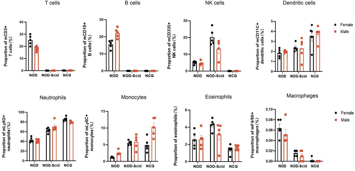
图1 NOD、NOD-Scid和NCG小鼠外周血免疫细胞比例的统计图(n=5)
7週齢のNOD、NOD-Scid、およびNCGマウスの周辺血液から採取され、その免疫細胞の割合を決定するためにフローアッセイが行われました。結果は、NODと比較して、NOD-ScidにはほとんどT細胞とB細胞が存在せず、NK細胞の割合が補償的に増加し、DC細胞、好中球、単球、および好酸球の割合が増加し、マクロファージの割合が減少したことを示しました。NODと比較して、NCGはほとんどT、B、NK細胞、およびマクロファージが存在せず、その免疫不全はNOD-Scidよりも完全であることが示されました。
2. NCG脾臓免疫細胞亜集団の統計
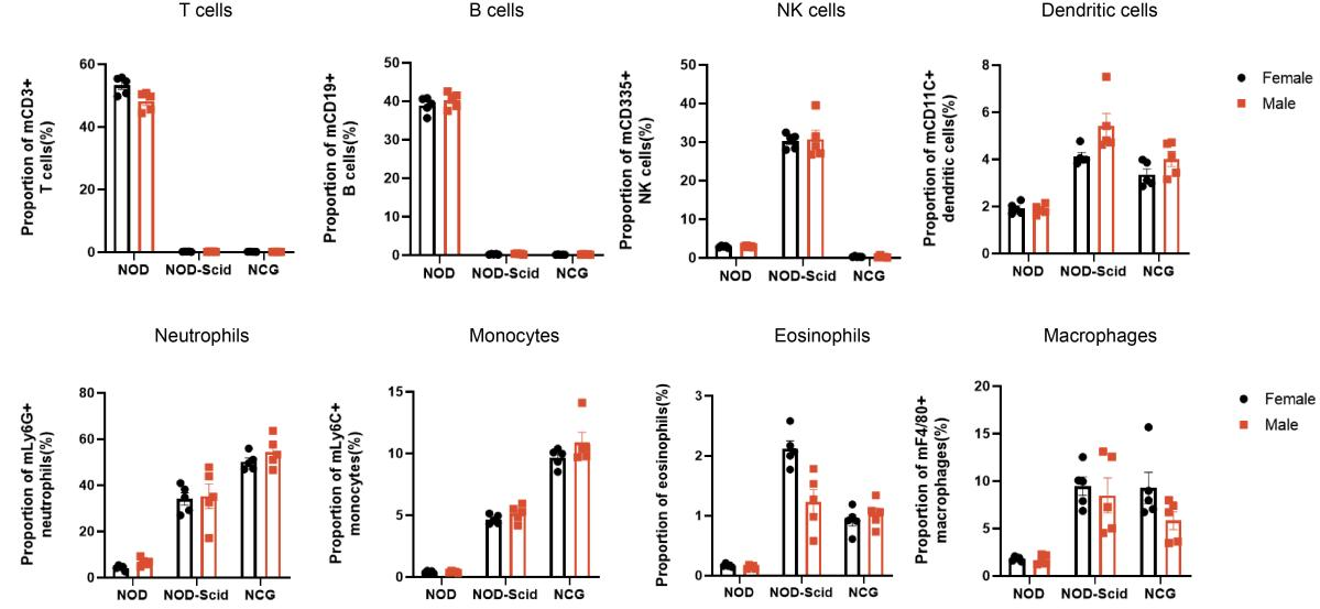
図2. NOD、NOD-Scid、NCGマウスの脾臓免疫細胞割合の統計プロット(n=5)
7週齢のNOD、NOD-Scid、およびNCGマウスの脾臓をフローアッセイのために採取し、その免疫細胞の分数比率を決定しました。結果は、NODと比較して、NOD-ScidにはほとんどT細胞とB細胞が存在せず、NK細胞の比率が補償的に増加し、DC細胞、好中球、単球、好酸球、およびマクロファージの比率が上昇していることを示しました。NODと比較して、NCGはほとんどT、B、およびNK細胞が存在せず、免疫不全の程度がより高いことが示されました。
3. NCG骨髄免疫細胞亜集団の統計
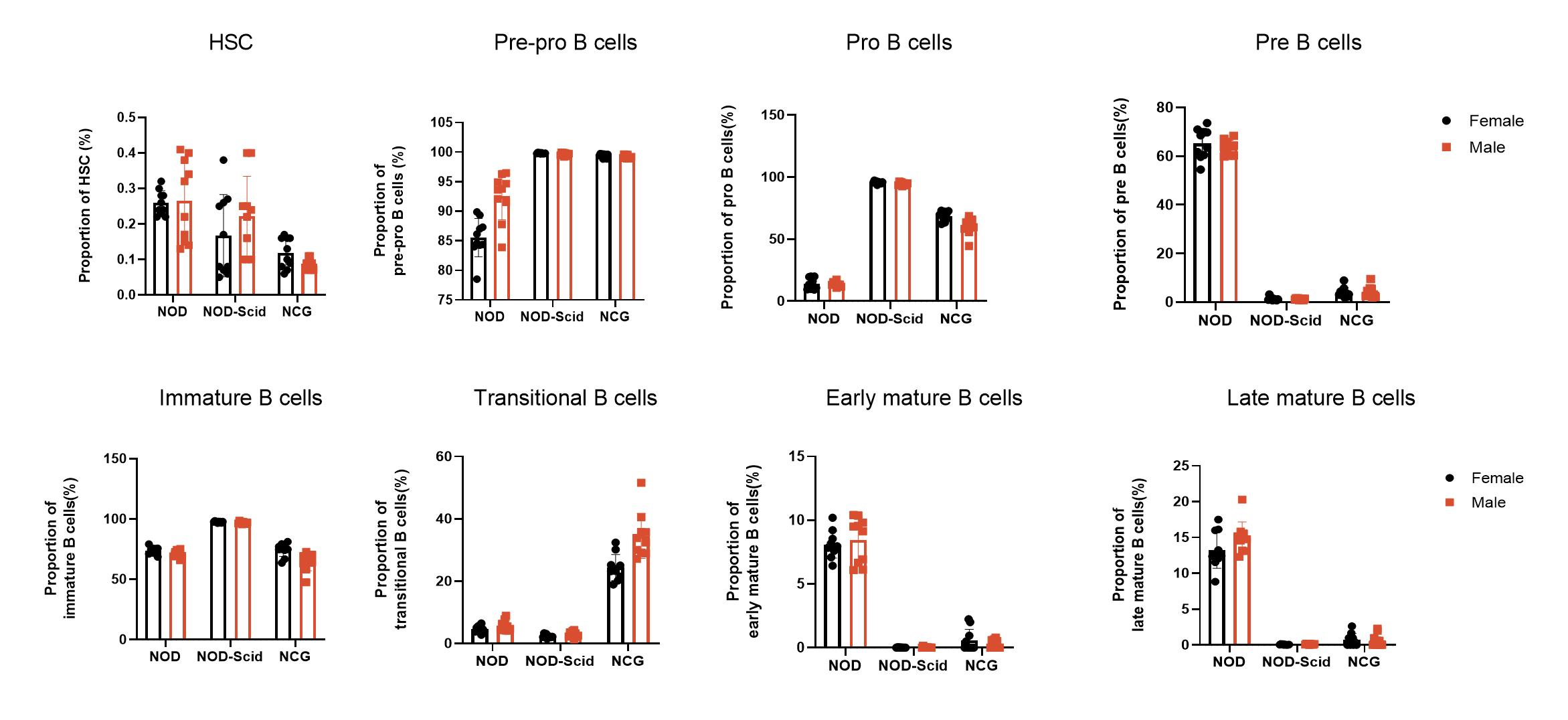
図3 NOD、NOD-Scid、NCGマウスの骨髄免疫細胞割合の統計プロット(n=5)
7週齢のNOD、NOD-Scid、およびNCGマウスの骨髄から、B細胞の生成中の各亜集団(造血幹細胞、Pre-pro-B、Pro-B、Pre-B、未成熟、移行期、初期Bおよび後期の成熟B細胞)の割合を決定するためにフローアッセイが行われました。結果は、NODと比較して、NOD-ScidおよびNCGマウスの骨髄においてPre-pro B細胞とPro B細胞が増加している一方、Pre-B細胞、早期・後期の成熟B細胞の含有量が減少していることを示しました。さらに、NCGマウスの骨髄において、移行期B細胞はNODおよびNOD-Scidマウスと比較して有意に増加していることが示されました。
4. NCG脾臓、胸腺、およびリンパ節のH&E染色
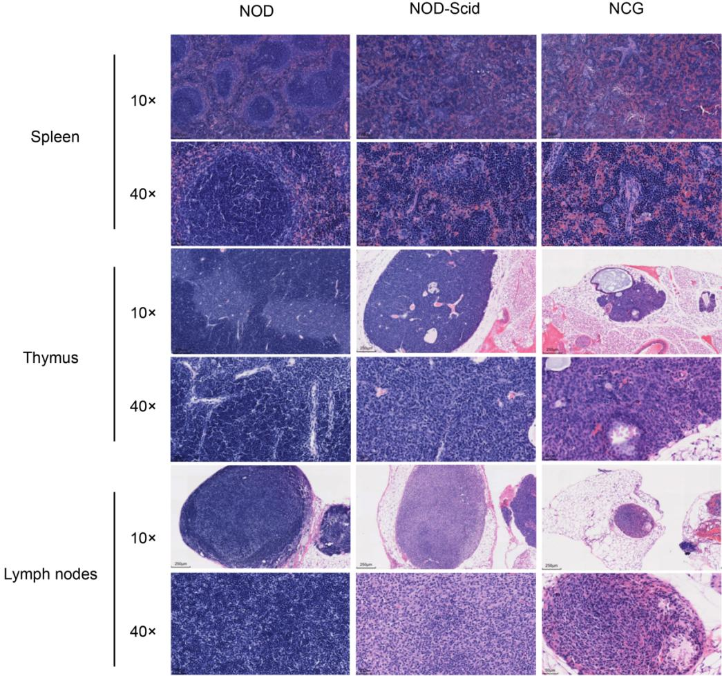
図4. NOD、NOD-Scid、NCGマウスの脾臓、胸腺、リンパ節のH&E染色結果
7週齢のNOD、NOD-Scid、およびNCGマウスの脾臓、胸腺、およびリンパ節は4% PFAで固定され、パラフィンに埋め込まれ、切片にされ、H&E染色が施され、その臓器構造に異常があるかどうかを確認するために行われました。結果は、NODマウスの脾臓には無傷の腹膜があり、肥厚は見られません。脾臓洞は拡張せず、脾臓小胞のサイズは異常に変化せず、赤髄と白髄の比率はほぼ正常でした。胸腺は無傷で、皮質と髄質は正常で、髄質に胸腺小胞が見られ、出血や萎縮などの病理学的変化は見られません。リンパ節皮質と髄質の比率は正常であり、リンパ球は減少せず壊死していません。一方、NOD-ScidおよびNCGマウスの脾臓リンパ節は小さくなり、リンパ球の数が減少します。胸腺の皮質と髄質は明確ではなく、胸腺は萎縮し、皮質および髄質のリンパ球が顕著に減少します。リンパ節皮質と髄質も明確ではなく、皮質が萎縮し、皮質および髄質のリンパ球が顕著に減少します。 H&E染色の結果は、体の視覚とフローサイトメトリーの結果と一致します。
さらに、7週齢のNOD、NOD-Scid、およびNCGマウスの心臓、肝臓、肺、腎臓、大腸、および睾丸(オス)はH&E染色のために取られました。結果は、これらの3つの系統が正常な臓器組織構造を持っていたことを示しました。
5. NCGの臓器指数およびリンパ節の数
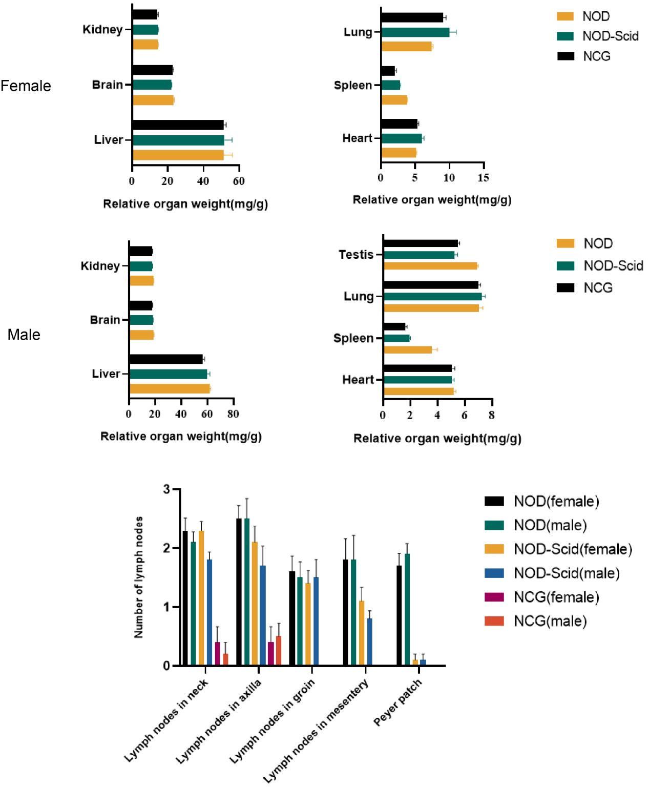
Figure 5. NOD, NOD-Scid and NCG mice comparison of organ indices and number of lymph nodes (n=10)
The hearts, livers, spleens, lungs, kidneys, brains and testes (males) of 7-week-old NOD, NOD-Scid and NCG mice were taken for organ weighing and organ indices were calculated. The results showed that the spleen index was significantly reduced in NOD-Scid and NCG mice compared to NOD mice, and the reduction was even more significant in NCG. The remaining organ indices were not significantly different.
In addition, by counting the cervical lymph nodes, axillary lymph nodes, inguinal lymph nodes, mesenteric lymph nodes and Paired lymph nodes in mice, the results showed that the number of lymph nodes was significantly reduced in NOD-Scid and NCG mice compared with NOD mice, while all NCG mice had no inguinal lymph nodes, mesenteric lymph nodes and Paired lymph nodes.
6. NCGの成長曲線
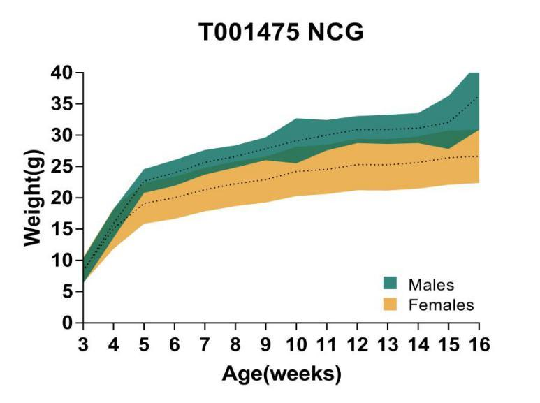
図6. NCGマウスの成長曲線
16週齢のNCGマウスに対して体重解析が行われました。結果から、NCGの雄マウスの体重は一般的に雌マウスよりも高かったことがわかってきました。
7. 通常の血液分析
NOD | NOD-Scid | NCG | ||||
Female(n=5) (Mean±SEM) | Male(n=5) (Mean±SEM) | Female(n=5) (Mean±SEM) | Male(n=5) (Mean±SEM) | Female(n=5) (Mean±SEM) | Male(n=5) (Mean±SEM) | |
WBC(K/uL) | 2.37±0.77 | 2.45±1.16 | 1.37±0.46 | 1.19±0.28 | 0.88±0.21 | 1.15±0.26 |
Lym(K/uL) | 1.35±0.38 | 1.24±0.70 | 0.26±0.08 | 0.30±0.06 | 0.12±0.04 | 0.29±0.15 |
Mon(K/uL) | 0.227±0.15 | 0.28±0.15 | 0.20±0.07 | 0.10±0.06 | 0.04±0.02 | 0.05±0.03 |
Neu (K/uL) | 0.67±0.29 | 0.81±0.35 | 0.79±0.32 | 0.72±0.20 | 0.66±0.19 | 0.76±0.13 |
Eos (K/uL) | 0.12±0.06 | 0.12±0.02 | 0.10±0.05 | 0.07±0.02 | 0.07±0.02 | 0.05±0.01 |
Bas (K/uL) | 0.01±0.01 | 0.01±0.01 | 0.01±0.01 | 0.003±0.006 | 0.00±0.00 | 0.00±0.00 |
RBC(M/uL) | 7.68±0.22 | 8.00±0.35 | 7.46±0.40 | 8.39±0.84 | 7.98±0.68 | 7.87±0.54 |
HGB(g/L) | 144.60±4.83 | 149.00±4.64 | 134.60±6.27 | 147.80±11.39 | 143.00±11.90 | 140.20±9.68 |
HCT(%) | 39.88±1.15 | 42.04±1.85 | 38.82±2.34 | 43.64±4.13 | 41.24±3.63 | 40.82±3.29 |
MCV (fL) | 51.96±0.53 | 52.62±0.62 | 52.10±0.66 | 52.12±0.62 | 51.74±0.35 | 51.90±0.85 |
MCH (pg) | 18.76±0.31 | 18.60±0.40 | 18.00±0.16 | 17.62±0.43 | 17.88±0.15 | 17.76±0.30 |
MCHC(g/L) | 362.00±4.95 | 354.20±5.98 | 346.40±6.07 | 338.80±8.64 | 346.60±3.78 | 343.20±5.54 |
RDW_CV(%) | 13.12±0.16 | 13.46±0.49 | 13.70±0.70 | 13.60±0.20 | 13.44±0.34 | 13.92±0.18 |
RDW_SD(fL) | 42.84±1.18 | 42.68±0.75 | 43.20±0.96 | 43.20±1.13 | 43.02±1.00 | 42.52±1.06 |
PLT(K/uL) | 1289.6±89.45 | 1482.60±175.24 | 1375.40±167.61 | 1533.60±236.50 | 1226.20±171.79 | 1665.60±45.92 |
MPV (fL) | 4.84±0.17 | 5.06±0.23 | 4.86±0.09 | 4.74±0.11 | 4.74±0.20 | 4.78±0.11 |
PDW(fL) | 10.62±0.57 | 11.42±0.86 | 10.64±0.36 | 10.62±0.43 | 10.54±0.75 | 10.64±0.22 |
PCT(%) | 0.62±0.05 | 0.74±0.07 | 0.66±0.09 | 0.72±0.12 | 0.58±0.06 | 0.79±0.03 |
P_LCR(%) | 19.82±1.73 | 22.62±3.13 | 20.85±1.18 | 19.86±1.72 | 18.77±2.63 | 19.33±1.48 |
P_LCC(K/uL) | 255.0±23.24 | 331.80±32.77 | 286.60±42.51 | 304.40±60.66 | 226.60±17.04 | 321.40±27.20 |
NRBC(K/uL) | 5.28±11.50 | 10.77±14.56 | 0.30±0.54 | 0.36±0.72 | 0.03±0.01 | 0.04±0.01 |
ALY(K/uL) | 0.03±0.008 | 0.03±0.02 | 0.001±0.002 | 0.003±0.005 | 0.00±0.00 | 0.01±0.01 |
LIC(K/uL) | 0.01±0.006 | 0.01±0.002 | 0.01±0.006 | 0.01±0.006 | 0.01±0.003 | 0.01±0.004 |
表1. 7週齢のNOD、NOD-Scid、およびNCGマウスにおける全血球数の解析
8. 血液生化学分析
NOD | NOD-Scid | NCG | ||||
Female(n=5) (Mean±SEM) | Male(n=5) (Mean±SEM) | Female(n=5) (Mean±SEM) | Male(n=5) (Mean±SEM) | Female(n=5) (Mean±SEM) | Male(n=5) (Mean±SEM) | |
ALT(IU/L) | 21.72±5.47 | 34.48±4.60 | 24.04±7.87 | 62.56±40.44 | 51.44±67.41 | 41.40±12.92 |
AST(IU/L) | 131.64±41.36 | 160.28±38.45 | 130.64±13.96 | 191.12±67.60 | 168.24±100.82 | 142.00±36.95 |
CK(mg/L) | 296.40±222.91 | 572.80±346.63 | 198.40±25.67 | 337.20±165.49 | 194.00±20.35 | 196.40±40.65 |
ALB(g/L) | 42.16±2.00 | 40.28±2.36 | 42.68±3.20 | 39.64±2.77 | 42.36±2.03 | 40.44±2.71 |
TBIL(umol/L) | 1.69±0.67 | 2.38±0.43 | 1.84±0.21 | 2.33±0.51 | 1.38±0.40 | 1.95±0.71 |
BUN(mmol/L) | 7.28±0.92 | 8.97±1.00 | 8.11±1.02 | 10.55±1.27 | 8.20±0.70 | 8.46±1.02 |
CREA(umol/L) | 7.40±3.72 | 6.36±2.45 | 5.96±1.04 | 8.12±1.15 | 6.40±1.92 | 6.76±2.30 |
CHO(mmol/L) | 2.68±0.17 | 3.88±0.44 | 2.47±0.31 | 3.06±0.47 | 2.32±0.30 | 3.58±0.31 |
TG(mmol/L) | 0.62±0.23 | 1.35±0.62 | 0.70±0.32 | 1.65±0.91 | 0.45±0.17 | 1.52±0.46 |
LDH(IU/L) | 2738.80±527.71 | 2986.40±579.96 | 3297.20±625.20 | 3470.00±554.64 | 3258.00±193.99 | 3557.2±559.94 |
LDL(mmol/L) | 0.29±0.05 | 0.26±0.01 | 0.25±0.09 | 0.08±0.04 | 0.30±0.08 | 0.31±0.07 |
GLU(mmol/L) | 1.52±0.74 | 1.44±0.87 | 1.02±1.06 | 3.36±2.04 | 0.64±0.67 | 2.26±1.78 |
HDL(mmol/L) | 2.06±0.14 | 3.09±0.34 | 1.86±0.24 | 2.42±0.37 | 1.71±0.23 | 2.78±0.22 |
TP(g/L) | 59.88±4.68 | 62.96±4.02 | 58.56±4.37 | 61.20±4.84 | 57.96±1.97 | 61.52±4.05 |
AKP(IU/L) | 213.20±10.35 | 182.40±10.71 | 184.80±21.48 | 160.00±10.49 | 205.60±7.54 | 150.40±14.99 |
表2. 7週齢のNOD、NOD-Scid、およびNCGマウスにおける血液生化学分析
9. NCGのがん原性試験
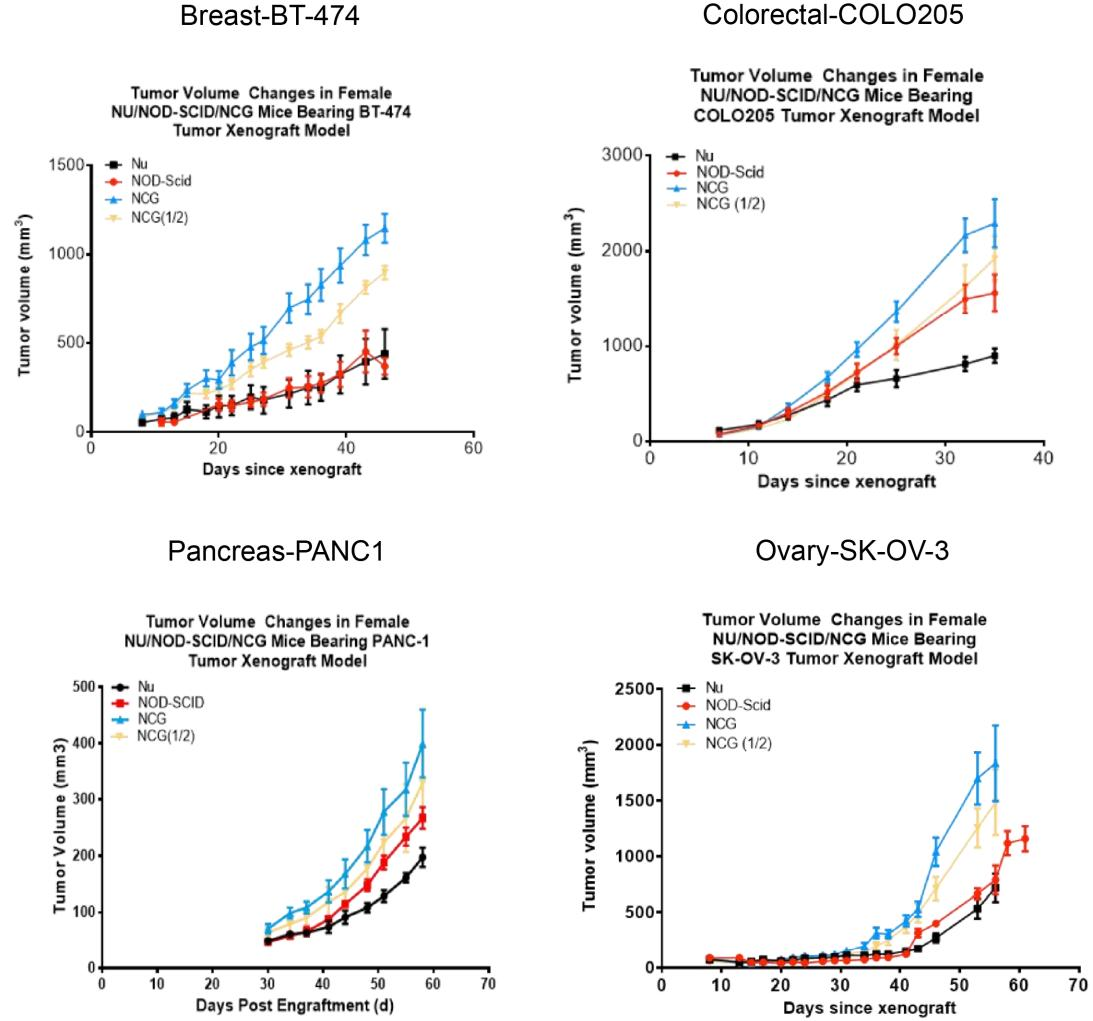
図7. NCGマウスの腫瘍形成性試験
ヒトの乳がん細胞株BT-474、ヒトの大腸がん細胞株COLO205、ヒトの膵臓がん細胞株PANC1、およびヒトの卵巣がん細胞株SK-OV-3は、それぞれ対数成長段階にある6~8週齢のNu/NOD-Scid/NCGマウスに皮下接種され、腫瘍は時間の経過とともに大きさに従った勾配曲線で成長しました(腫瘍体積の値は平均±SEMで表現されています)。
10. NCG効果試験
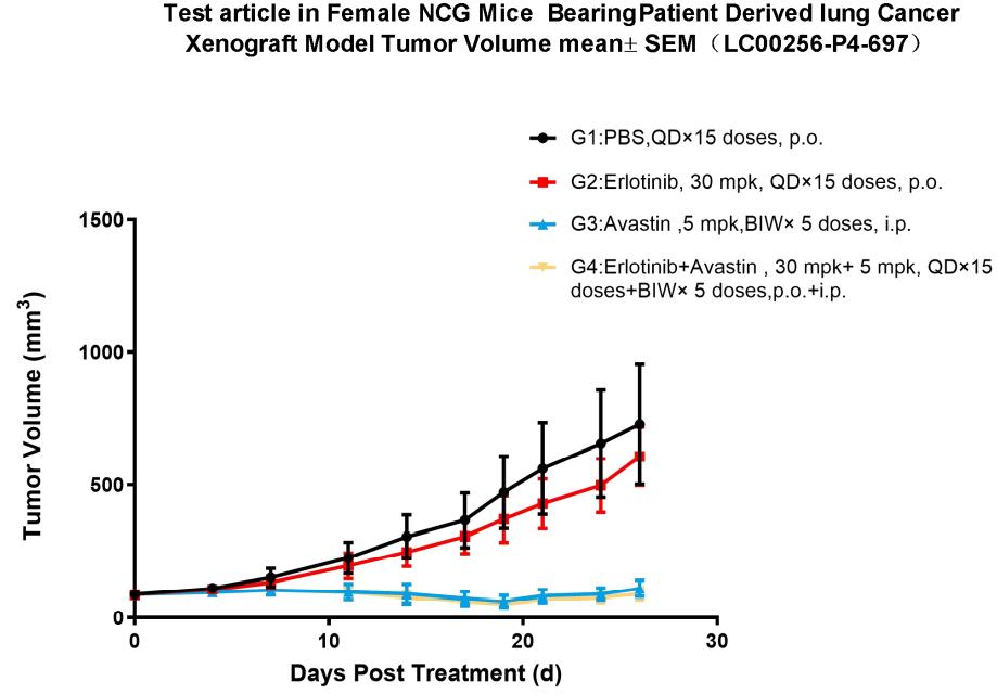
図8. 肺癌のPDXモデルに皮下移植されたNCGマウスでのエルロチニブの薬力学的評価
NCGマウスにおける皮下移植肺がんPDXモデルを構築し、エルロチニブの抗腫瘍効果を評価しました。結果から、エルロチニブ処理群はモデル対照群と比較して腫瘍の成長を有意に抑制できます。さらに、アバスチンとの組み合わせは、エルロチニブ単独群と比較して腫瘍の成長を有意に抑制しました(腫瘍体積の値は平均±SEMで表現されています)。
公開文献
Hu B, Yu M, Ma X, et al. Interferon-a potentiates anti-PD-1 efficacy by remodeling glucose metabolism in the hepatocellular carcinoma microenvironment. Cancer discovery. Apr 12 2022;doi:10.1158/2159-8290.Cd-21-1022. (IF:39.397)
Ma W, Yang Y, Zhu J, et al. Biomimetic Nanoerythrosome‐Coated Aptamer‐DNA Tetrahedron/Maytansine Conjugates: pH‐Responsive and Targeted Cytotoxicity for HER2‐positive Breast Cancer. Advanced Materials.2109609. (IF:30.849)
Song H, Liu D, Wang L, et al. Methyltransferase like 7B is a potential therapeutic target for reversing EGFR-TKIs resistance in lung adenocarcinoma. Molecular cancer. Feb 10 2022;21(1):43. (IF:41.444)
Zhang L, Zhu Z, Yan H, et al. Creatine promotes cancer metastasis through activation of Smad2/3. Cell metabolism. 2021;33(6):1111-1123. e4. (IF:22.4)
Liu C, Zou W, Nie D, et al. Loss of PRMT7 reprograms glycine metabolism to selectively eradicate leukemia stem cells in CML. Cell Metab. Apr 26 2022. (IF:22.4)
Zhang X-N, Yang K-D, Chen C, et al. Pericytes augment glioblastoma cell resistance to temozolomide through CCL5-CCR5 paracrine signaling. Cell Research. 2021:1-16. (IF:25.617)
Dai Z, Mu W, Zhao Y, et al. T cells expressing CD5/CD7 bispecific chimeric antigen receptors with fully human heavy-chain-only domains mitigate tumor antigen escape. Signal transduction and targeted therapy. Mar 25 2022;7(1):85. (IF:18.187)
Dai Z, Liu H, Liao J, et al. N7-Methylguanosine tRNA modification enhances oncogenic mRNA translation and promotes intrahepatic cholangiocarcinoma progression. Molecular Cell. 2021. (IF:17.97)
Hao M, Hou S, Li W, et al. Combination of metabolic intervention and T cell therapy enhances solid tumor immunotherapy. Science Translational Medicine. 2020;12(571). (IF:17.956)
Liu Y, Liu G, Wang J, et al. Chimeric STAR receptors using TCR machinery mediate robust responses against solid tumors. Science Translational Medicine. 2021;13(586). (IF:17.956)
Yan H, Wang Z, Sun Y, Hu L, Bu P. Cytoplasmic NEAT1 Suppresses AML Stem Cell Self‐Renewal and Leukemogenesis through Inactivation of Wnt Signaling. Advanced Science. 2021;8(22):2100914. (IF:17.521)
Luo Q, Wu X, Chang W, et al. ARID1A prevents squamous cell carcinoma initiation and chemoresistance by antagonizing pRb/E2F1/c-Myc-mediated cancer stemness. Cell Death & Differentiation. 2020;27(6):1981-1997. (IF:12.067)
Wu M, Zhang X, Zhang W, et al. Cancer stem cell regulated phenotypic plasticity protects metastasized cancer cells from ferroptosis. Nature communications. Mar 16 2022;13(1):1371. (IF:14.919)
Liu H, Bai L, Huang L, et al. Bispecific antibody targeting TROP2xCD3 suppresses tumor growth of triple negative breast cancer. Journal for ImmunoTherapy of Cancer. 2021;9(10):e003468. (IF:12.469)

